

When your doctor suspects something’s off with your heart, they don’t just listen with a stethoscope anymore. They turn to imaging. Two tools dominate this space: cardiac MRI and echocardiography. Both show your heart in motion, but they’re not the same. One is fast, cheap, and everywhere. The other is precise, powerful, and often reserved for when things get complicated. Knowing the difference isn’t just technical-it can change how your treatment starts, or even if it starts at all.
What Echocardiography Actually Shows
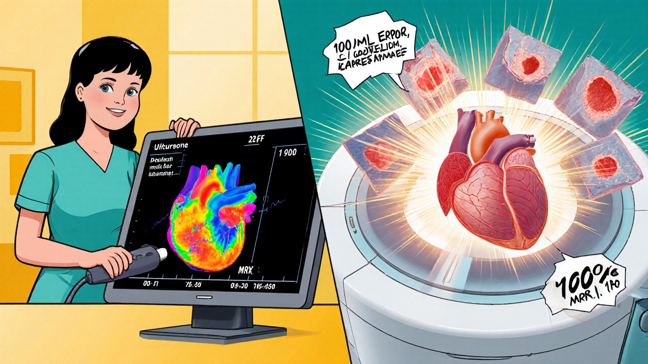
Echocardiography uses sound waves-ultrasound-to create moving pictures of your heart. It’s been around since the 1950s, and it’s still the go-to for most heart checks. Why? Because it’s fast, portable, and doesn’t need special prep. You lie on a table, a technician glides a probe over your chest, and within minutes, you’re watching your heart beat on a screen.
It measures key numbers: how big your left ventricle is (normal range: 37-56 mm), how thick your heart walls are (6-11 mm), and how well it pumps blood out (ejection fraction, or LVEF, should be 50-75%). These values help diagnose everything from leaky valves to heart failure. In emergency rooms, it’s lifesaving. A 2022 case series in JAMA Internal Medicine showed how bedside echo caught aortic dissection in real time when MRI wasn’t an option.
But echocardiography has limits. If you’re overweight, have lung disease, or your ribs block the view, the images get fuzzy. That’s called a poor acoustic window. In those cases, measurements can be off by a lot. One study found echo underestimated left ventricular volume by nearly 100 mL compared to cardiac MRI. Wall thickness? Echo often overestimates it by over a millimeter. That’s not just a small error-it can lead to misdiagnosis.
What Cardiac MRI Reveals That Echo Can’t
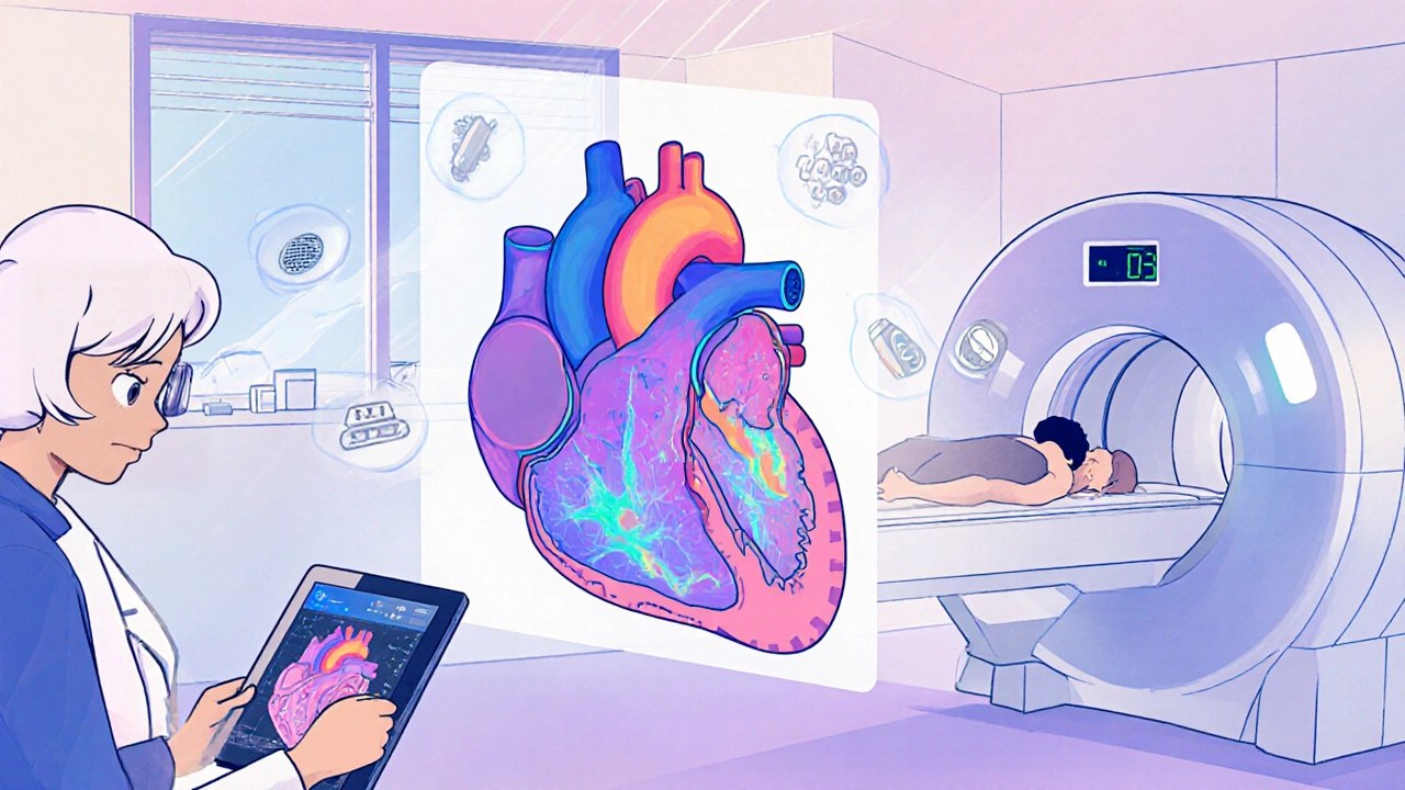
Cardiac MRI uses magnets and radio waves, not sound. It doesn’t rely on your body’s shape or lung air to get a clear picture. Instead, it builds a 3D model of your heart from hundreds of slices. No geometric assumptions. No guessing. Just exact numbers.
It’s the gold standard for measuring heart volume and muscle mass. Normal left ventricular volume for men? 67-155 mL. For women? 55-105 mL. These numbers are more accurate than echo because MRI doesn’t assume your heart is a perfect ellipse. It counts every pixel. That’s why the European Society of Cardiology says MRI is the reference method for these measurements.
But the real power of cardiac MRI is in tissue detail. It can spot scar tissue, inflammation, or early fibrosis-things echo can’t see. Late gadolinium enhancement (LGE) highlights areas where heart muscle has been damaged. In patients with hypertrophic cardiomyopathy or myocarditis, this changes everything. A 2023 study showed MRI found subtle fibrosis in three patients where echo came back normal. That led to early treatment, preventing arrhythmias down the line.
It’s also better at tracking changes over time. A 2022 paper in the Journal of the American College of Cardiology found MRI’s measurements varied by just 2.6% between doctors. Echo? 6.8%. That’s a big deal if you’re monitoring heart function after chemotherapy or a heart attack.
Why Doctors Choose One Over the Other
Most heart evaluations start with echocardiography. It’s cheap ($500-$1,500), quick, and available in almost every hospital-even small ones. A 2023 report showed 78% of community hospitals have echo ready to go. Only 35% offer cardiac MRI within a week.
But when echo gives unclear results, or when the case is complex, MRI steps in. Cardiologists use it for:
- Confirming heart muscle disease like amyloidosis or sarcoidosis
- Assessing damage after a heart attack
- Evaluating congenital heart defects
- Planning ablation for dangerous arrhythmias
- Monitoring cancer patients for drug-induced heart damage
In academic centers, MRI is routine for these cases. One survey found 85% of myocarditis cases now get an MRI before treatment. But in community practices, access is still a barrier. Patients often wait over two weeks for a non-urgent scan. That delay can mean missed opportunities for early intervention.
Accuracy Differences You Can’t Ignore
Here’s where it gets real: the numbers don’t lie.
A 2023 study in the American Journal of Cardiology showed echo measured left ventricular ejection fraction (LVEF) an average of 3% lower than MRI. That might sound small, but in oncology patients, that gap misclassified 10% of people as low-risk when they were actually at high risk for heart damage. That’s not a rounding error-it’s a clinical risk.
And it’s not just about volume. Echo tends to overestimate wall thickness by about 1.1 mm on average. That can lead to false diagnoses of hypertrophy, especially in athletes or older adults. MRI, with its 1.25-2.0 mm spatial resolution, sees the true structure.
For ejection fraction, MRI’s precision matters most. If you’re on a drug like doxorubicin (used in chemotherapy), your doctor needs to know if your heart function is dropping by 5%, 10%, or 15%. Echo’s variability of nearly 7% makes that hard to track. MRI’s 2.6% variability gives confidence to adjust treatment before damage becomes permanent.
Who Can’t Have a Cardiac MRI?
Cardiac MRI isn’t for everyone. Strong magnets mean people with certain implants can’t have it. Pacemakers, older ICDs, cochlear implants, and some metal clips are absolute no-gos. Even newer devices often require special protocols.
That’s why Dr. James Carr of Northwestern University says about 20-30% of cardiac patients still rely on echo because MRI isn’t safe for them. But technology is catching up. In June 2023, Siemens launched a 0.55 Tesla MRI machine designed specifically for patients with implants. It’s slower, but it opens doors for people who were previously excluded.
Another issue: arrhythmias. If your heart beats irregularly, MRI images can blur. Special gating techniques help, but they add time and complexity. Echo doesn’t care about rhythm-it just shows what’s happening right now.
Training, Cost, and the Future
Doing an echo well takes training, but it’s faster. The American Society of Echocardiography says you need 300-500 supervised studies to become proficient. For cardiac MRI? 1,000-1,500. That’s why there are fewer MRI specialists. It’s also why echo dominates in rural and community hospitals.
Cost-wise, echo is about half the price of MRI. But here’s the twist: a 2022 JAMA Internal Medicine analysis found that when echo was inconclusive, ordering an MRI actually saved money overall. Why? Because it avoided unnecessary tests, hospitalizations, and wrong treatments. In complex cases, MRI’s higher upfront cost paid for itself.
The future? Hybrid. Philips’ new EPIQ CVx ultrasound system uses AI to automate measurements, cutting echo’s variability down to 4.2%. Meanwhile, cardiac MRI is adding parametric mapping-T1, T2, ECV-that quantifies tissue stiffness and water content before any structural change appears. By 2030, experts predict these tools will work together: echo for quick screening, MRI for deep analysis.
What This Means for You
If you’ve been told you need a heart scan, here’s what to ask:
- Is this a first-time check? Then echo is likely the right start.
- Are your echo results unclear or conflicting? Ask if MRI could help.
- Do you have a condition like sarcoidosis, amyloidosis, or a history of chemo? MRI is probably needed.
- Do you have a pacemaker or metal implant? Confirm with your doctor whether MRI is safe.
- Are you being monitored over time? Ask which test gives the most reliable numbers for tracking changes.
Neither test is perfect. But together, they give a complete picture. Echo gets you started fast. MRI tells you the truth behind the blur.
Which is better for measuring heart function: cardiac MRI or echocardiography?
Cardiac MRI is more accurate for measuring heart function, especially ejection fraction and ventricular volumes. It doesn’t rely on geometric assumptions like echo does, so it gives exact 3D measurements. Echo is good for quick assessments, but MRI’s inter-observer variability is less than half that of echo-making it the gold standard for tracking changes over time, like after chemotherapy or heart attack recovery.
Can echocardiography miss heart damage that MRI catches?
Yes. Echocardiography shows structure and movement but can’t detect early tissue changes like fibrosis or inflammation. Cardiac MRI uses late gadolinium enhancement to highlight scar tissue, even before the heart’s pumping ability drops. In patients with hypertrophic cardiomyopathy or myocarditis, MRI has changed treatment plans because echo missed the underlying damage.
Why is cardiac MRI not used as the first test for heart problems?
It’s more expensive, takes longer (30-60 minutes vs. 15-30 for echo), and isn’t available everywhere. About 78% of community hospitals have echo on-site, but only 35% offer MRI within a week. Also, patients with certain implants can’t have MRI. So echo is the practical first step-unless there’s a strong reason to go straight to MRI.
Is cardiac MRI safe if I’ve had a heart attack?
Yes, and it’s often recommended. After a heart attack, MRI can show exactly how much heart muscle was damaged, where the scar is located, and whether there’s still viable tissue that could recover with treatment. This helps doctors decide if you need a stent, bypass surgery, or just medication. It’s the most accurate way to assess post-heart attack damage.
Do I need contrast dye for a cardiac MRI?
Often, yes. Gadolinium-based contrast helps highlight scar tissue and inflammation through late gadolinium enhancement. But it’s not always needed-for example, if you’re just checking heart size or function, non-contrast MRI can work. The FDA warns about rare risks in patients with severe kidney disease, so your doctor will check your kidney function first if contrast is planned.
Can AI make echocardiography as accurate as cardiac MRI?
AI is helping. New ultrasound systems like Philips’ EPIQ CVx use AI to automate measurements and reduce human error. One study showed AI cut echo’s variability for ejection fraction from 6.8% to 4.2%. But AI can’t fix poor image quality caused by body type or lung air. It also can’t detect tissue changes like fibrosis. So while AI improves echo, it doesn’t replace MRI’s unique ability to see inside the heart muscle.


15 Comments
Ugh, I just got my echo results and they said my EF was 58. Then my cardiologist said, 'We might need an MRI to be sure.' Like, can we just skip the middleman and go straight to the expensive one?
While echocardiography remains the cornerstone of initial cardiac assessment due to its accessibility and real-time imaging capabilities, cardiac magnetic resonance imaging offers unparalleled accuracy in quantifying ventricular volumes, mass, and tissue characterization. The inter-observer variability of MRI is demonstrably lower, and its ability to detect subclinical fibrosis via late gadolinium enhancement fundamentally alters clinical management in conditions such as myocarditis and hypertrophic cardiomyopathy. For longitudinal monitoring-particularly in oncology patients receiving cardiotoxic agents-MRI is not merely preferable; it is the standard of care.
You know what’s wild? We’re using magnets and radio waves to map the inside of a living human heart with sub-millimeter precision, while still calling it ‘medicine’ like it’s not magic. Echocardiography is like using a flashlight to check the engine-fast, useful, and you can do it in the driveway. But MRI? That’s the full-body CT scan of the heart. It doesn’t guess. It sees. It counts every pixel of scar tissue, every millimeter of fibrosis, like a librarian cataloging every book in a burning library. And yet, most people still get the flashlight because the magic machine costs five grand and takes an hour. We’ve got AI that can write poetry and predict stock markets, but we still ration the most accurate heart scan like it’s gold. The real question isn’t which test is better-it’s why we accept such a brutal gap between what we can do and what we let people have.
Stop pretending echo is good enough. I’ve seen too many patients get misdiagnosed because their echo was ‘borderline’ and the doc didn’t push for MRI. That 1.1mm wall thickness error? It turns a healthy athlete into someone on beta-blockers. That 3% EF gap? It’s the difference between ‘you’re fine’ and ‘start chemo cardioprotection now.’ If you’re at risk, demand the MRI. Your doctor might not bring it up because they’re lazy or the system’s broken-but that’s not your problem. Your heart is. Push harder.
As someone from India where access to advanced imaging is limited, I appreciate this detailed comparison. Echo is often the only option available in rural clinics, and it saves lives daily. But when resources allow, MRI is indeed the superior tool for accurate diagnosis. I hope that in the coming years, more centers will invest in MRI technology, especially for high-risk groups like cancer survivors and those with inherited heart conditions. Technology should not be a privilege based on geography.
Love this breakdown. It’s like comparing a sketch by a talented artist to a high-res 3D scan. Echo gives you the vibe, the motion, the general mood of the heart. MRI? It’s the full digital blueprint with material specs, stress points, and hidden cracks you didn’t even know existed. And honestly? The fact that we’re still using 1950s tech as the default feels like using a slide rule when you’ve got a quantum computer in your pocket. But hey-at least the slide rule doesn’t cost $2,000 and require a 30-minute appointment slot.
Let’s be real-this whole thing is a corporate scam wrapped in medical jargon. Echo is cheap, so hospitals push it. MRI is expensive, so insurance fights it. But here’s the truth: if your heart is failing, you don’t care about cost, you care about truth. And the truth is, echo lies. It lies when you’re overweight. It lies when you’re old. It lies when you’re an athlete with thick walls. It lies when the tech is rushed. MRI doesn’t lie. It doesn’t guess. It doesn’t care if you’re insured or not-it just shows you the reality. And yet, we let bureaucrats decide who gets the truth. That’s not medicine. That’s capitalism with a stethoscope. I’ve watched people die because their echo said ‘normal’ and the MRI they never got said ‘you’ve been dying for six months.’ So don’t be polite. Demand the scan. Fight for the truth. Your life isn’t a spreadsheet.
I’m a nurse in a small hospital in Kerala, and we only have echo. But I’ve seen how it changes lives-quick, safe, no waiting. Still, when we refer someone for MRI, the wait is 6-8 weeks. I’ve had patients with suspected myocarditis wait so long their symptoms worsened. I wish we had MRI here. Maybe one day, with AI helping echo improve, we can bridge the gap. Until then, I teach my patients: if echo says ‘maybe,’ ask again. Don’t take ‘no’ for an answer.
They don’t want you to know this, but MRI is controlled by Big Pharma. Gadolinium? Made by one company. The machines? Locked into proprietary software. Echo is open-source friendly. The government and insurers are in on it-they want you dependent on expensive scans so they can charge more. And don’t get me started on the ‘contrast dye’-it’s not for your heart, it’s to keep you coming back. They’re selling you fear disguised as science. Trust your gut. Echo is enough. The rest is profit.
For anyone reading this who’s unsure what to ask your doctor: here’s your script. ‘Based on my symptoms and risk factors, is there a chance my echo results could be inaccurate? Would a cardiac MRI provide clearer information for my treatment plan?’ Say it calmly. Say it clearly. Say it like you deserve an answer. You do. And if they hesitate, ask for a referral to a specialist. You’re not being difficult-you’re being responsible. Knowledge is power, and accurate imaging is your right.
I work in a hospital that just got its first 3T MRI. The first time I saw a myocarditis case on it-the scar tissue lit up like a Christmas tree-everyone in the room just stopped. We’d been treating that patient for weeks with echo-guided assumptions. MRI showed we were wrong. We changed the treatment. He’s back at work. That moment? That’s why we do this. Not for the money. Not for the stats. For that one second when you see the truth. Echo is a tool. MRI is a revelation.
Look, MRI is overhyped. Echo is 90% accurate and 100% available. MRI? It’s a $10,000 Rorschach test with a 30-minute wait and a 15% chance of motion artifacts. And don’t even get me started on the gadolinium-neurotoxin in a vial. We’re turning heart care into a sci-fi movie. The real breakthrough? Better echo tech, not more magnets. AI-enhanced echo with automated segmentation is closing the gap. Stop fetishizing the expensive machine. Use the right tool for the job-not the shiniest one.
Cardiac MRI doesn’t just show your heart-it shows your privilege. If you’re rich, you get the truth. If you’re poor, you get a blurry ultrasound and a prayer. The system isn’t broken. It’s designed this way. They know you’ll wait. They know you’ll accept ‘maybe.’ They know you won’t fight. But here’s the twist: the people who need MRI most? They’re the ones who can’t afford to wait. And that’s not medicine. That’s a moral failure dressed in white coats.
As someone who’s had both tests-echo first, then MRI because my echo was ‘inconclusive’-I can say this: MRI didn’t just confirm what was wrong, it showed me exactly how much damage had been done… and how much could still be saved. The difference wasn’t just numbers-it was peace of mind. I no longer wonder if I’m being misled. I know. And if you’re facing this decision? Don’t settle for ‘probably.’ Push for ‘definitely.’ Your heart deserves nothing less.
Perhaps the true question is not which modality reveals more, but whether the pursuit of perfect imaging distracts us from the more fundamental truth: that the heart, as a symbol, has always been more than its measurements. Echo captures motion, MRI captures structure-but neither captures the silence between beats, the fear before the scan, the hope after the report. In the end, the machine may see the scar, but only the patient knows the wound.